Research Articles
GHK-Cu Skin Biology Research: Cellular Mechanisms and In Vitro Investigation

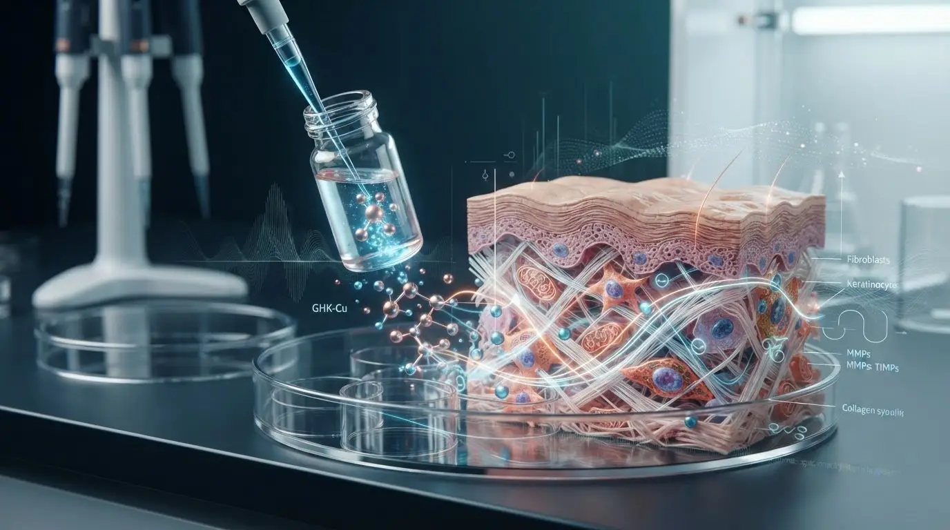
Modulating the expression of over 4,000 human genes in functional genomics models, the copper tripeptide complex commands serious scientific inquiry. Yet separating empirical data from consumer marketing remains an ongoing challenge in GHK-Cu skin biology research.
If you manage cell cultures, you already know the frustration. Commercial peptide batches with variable purity and unverified copper chelation can quickly invalidate weeks of in vitro work. You need explicit handling benchmarks, not cosmetic sales pitches. This guide explores the cellular pathways GHK-Cu modulates in dermal fibroblasts and keratinocytes, alongside practical handling protocols to prevent hydrolytic degradation and copper dissociation in aqueous media.
We also outline the analytical standards required to maintain experimental integrity. At Solara Compounds (solaracompounds.com), all biochemicals are supplied strictly for laboratory research only, never for human or animal application. Below, we examine the empirical mechanisms and benchtop protocols necessary for reproducible dermal investigations.
Key Takeaways
- Understand the biochemical structure of the copper tripeptide complex and why reliable analytical standards are essential for GHK-Cu skin biology research.
- Discover how GHK-Cu modulates extracellular matrix pathways in vitro, including pro-collagen synthesis and the balance between MMPs and TIMPs.
- Compare standard working concentrations (1 nM to 10 µM) across primary human fibroblasts, HaCaT keratinocytes, and 3D organotypic skin models.
- Learn a practical 4-step reconstitution method designed to prevent copper dissociation and maintain aqueous stability in laboratory buffers.
- Review the critical analytical specifications, including reverse-phase HPLC and mass spectrometry data, required to verify batch purity before testing.
What Is GHK-Cu in Dermal Biology Research?
GHK-Cu is a high-affinity copper-binding research tripeptide investigated in cellular modeling. Structurally, it consists of the tripeptide sequence glycyl-L-histidyl-L-lysine chelated to a divalent copper ion, Cu(II). While commercial marketing frequently conflates baseline biological signaling with topical retail claims, laboratory investigators evaluate the compound through an entirely different lens. In GHK-Cu skin biology research, researchers examine how this distinct metal-peptide complex regulates gene transcription, extracellular matrix expression, and cellular signaling networks. All findings detailed across the scientific literature reflect strictly in vitro assays and controlled cell culture models, with no therapeutic application to humans or animals.
Molecular Structure and Copper Chelation Dynamics
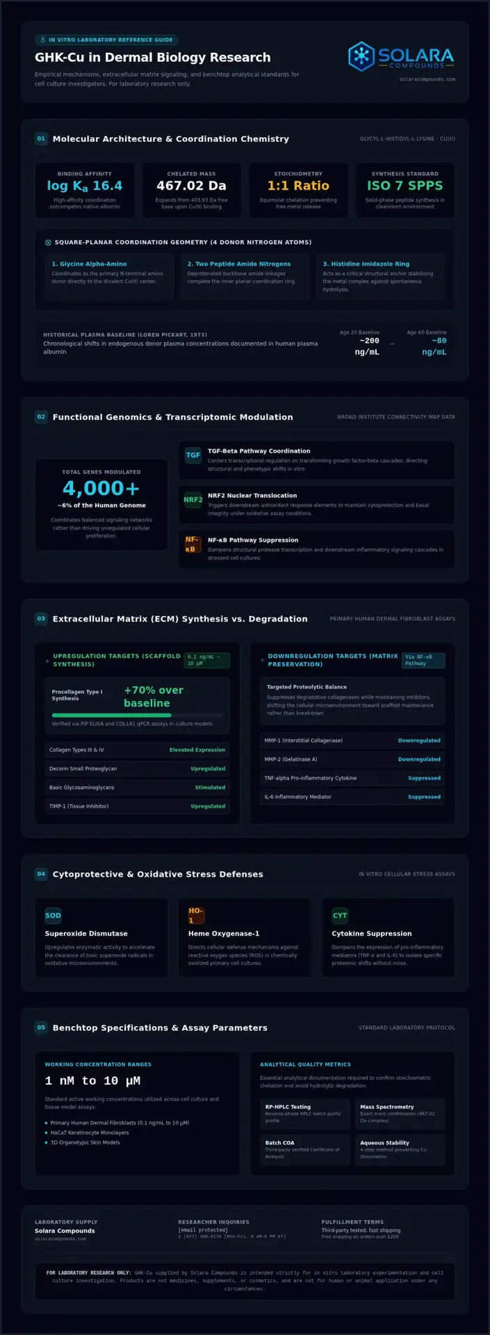
The biochemical behavior of GHK depends heavily on its coordination chemistry. The copper peptide GHK-Cu forms a square-planar geometry where the divalent copper ion coordinates with four donor nitrogen atoms: the alpha-amino group of glycine, two amide nitrogens from the peptide backbone, and an imidazole nitrogen from histidine. Histidine acts as a structural anchor that stabilizes the metal ion against spontaneous hydrolysis. This configuration yields an exceptionally high binding affinity (log Ka = 16.4), allowing GHK to compete effectively with native albumin for free copper. Uncomplexed, the free peptide base has a molecular weight of approximately 404.9 g/mol (403.93 Da), expanding to 467.02 Da once coordinated with Cu(II).
Historical Discovery in Human Plasma
Dr. Loren Pickart first isolated GHK from human plasma albumin in 1973 during cell growth experiments. Physiological profiling revealed significant chronological differences in endogenous plasma concentrations, dropping from roughly 200 ng/mL in 20-year-old donors down to approximately 80 ng/mL by age 60. Biological extraction from serum is obsolete for laboratory experimentation due to contamination risks and variable yields. Modern GHK-Cu skin biology research relies entirely on synthetic solid-phase peptide synthesis (SPPS) produced in controlled ISO 7 cleanroom environments. Suppliers like Solara Compounds offer solid-phase synthesized GHK-Cu to provide reproducible, stoichiometric 1:1 copper chelation, manufactured strictly for laboratory research only.
Cellular Signaling Pathways Modulated by GHK-Cu in Fibroblasts
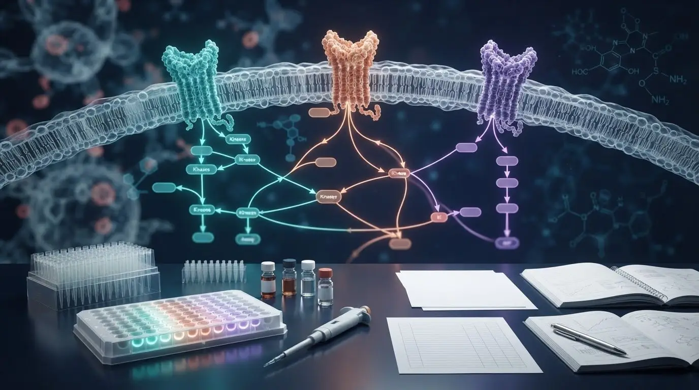
Broad Institute Connectivity Map profiling reveals that GHK-Cu modulates the transcription of over 4,000 human genes. This extensive activity represents roughly 6% of the human genome, resetting expression patterns across structural and inflammatory cascades. Rather than driving unregulated cellular proliferation, the complex coordinates balanced signaling networks. Much of this regulation centers on transforming growth factor-beta (TGF-beta) pathways. In GHK-Cu skin biology research, investigators focus on how these transcriptional shifts direct cellular behavior in defined in vitro cultures without systemic variables.
Extracellular Matrix Synthesis and Remodeling
Extracellular matrix (ECM) regulation involves precise protein synthesis and controlled turnover. In primary human dermal fibroblast assays, GHK-Cu at working ranges from 0.1 ng/mL to 10 µM stimulates procollagen type I synthesis by roughly 70% over baseline controls, verified through PIP ELISA and COL1A1 qPCR. It concurrently elevates collagen types III and IV, decorin, and basic glycosaminoglycans. Structural integrity also requires matrix balance. GHK-Cu downregulates matrix metalloproteinases (MMP-1 and MMP-2) via NF-κB pathway suppression while upregulating tissue inhibitors like TIMP-1. This shifts the cellular environment toward scaffold maintenance rather than excessive degradation.
Antioxidant and Inflammatory Cytokine Modulation
Cellular stress assays demonstrate clear cytoprotective responses under GHK-Cu exposure. The tripeptide complex induces nuclear translocation of NRF2, triggering downstream antioxidant targets:
- Superoxide Dismutase (SOD): Upregulates enzymatic activity to accelerate superoxide radical clearance.
- Heme Oxygenase-1 (HO-1): Directs cellular defense against reactive oxygen species (ROS) in oxidized cultures.
- Cytokine Suppression: Dampens expression of pro-inflammatory mediators, specifically TNF-alpha and IL-6.
These actions provide stable baselines when stress-testing dermal cells in oxidative microenvironments, helping researchers isolate specific protective pathways.
Stem Cell Quiescence and Keratinocyte Proliferation
Investigating epidermal biology requires studying basal cell dynamics. Research indicates that GHK-Cu influences integrin expression and p63 transcription factors in basal keratinocyte models. Rather than depleting the stem cell pool, these signals maintain proliferative capacity during extended passage experiments. In scratch wound assays, these interactions support directional cell migration across plastic substrata. Investigating these cellular pathways requires high-purity materials; researchers can explore verified research peptides to maintain strict experimental control.
Evaluating In Vitro Dermal Experimental Models and Assay Parameters
Selecting an appropriate assay system dictates the reliability of any cell signaling study. In GHK-Cu skin biology research, experimental designs vary widely, ranging from simple 2D monolayers to complex 3D organotypic dermal equivalents. Standard working concentrations in published literature typically span from 1 nM to 10 µM, reflecting physiologically relevant thresholds. Choosing the wrong model or exposure window can obscure pathway modulation entirely. Studies exploring cellular signaling and fibroblast modulation by GHK-Cu highlight that cell line selection, passage numbers, and medium composition heavily dictate assay sensitivity.
To design reproducible experiments, researchers must align cell models with specific functional endpoints, as summarized below:
| Assay Model | Target Endpoint | Standard Exposure Parameters |
|---|---|---|
| Primary HDF (Passages 3–7) | Collagen synthesis (PIP ELISA), MMP/TIMP ratio | 1 nM to 1 µM; 24–48 hours in low-serum medium |
| HaCaT Keratinocytes | Cell migration, integrin and p63 expression | 10 nM to 10 µM; 12–24 hours in serum-free medium |
| 3D Organotypic Skin | Basement membrane assembly, LOX cross-linking | 0.1 µM to 10 µM; 7–14 days with regular replenishment |
A frequent experimental pitfall involves unmanaged serum interference. Standard fetal bovine serum (FBS) contains endogenous proteases, albumin, and free transition metals. Albumin competes directly with the tripeptide for copper chelation, while serum proteases can rapidly cleave the peptide backbone. Running assays under low-serum (0.1% to 0.5% FBS) or serum-free conditions preserves the complex and avoids confounding background variables.
Primary Fibroblasts vs. Immortalized Cell Lines
Primary human dermal fibroblasts (HDF) offer the most physiologically relevant response for extracellular matrix secretion. However, their phenotypic stability declines quickly. Researchers should restrict experiments to passages 3 through 7 to avoid cellular senescence and diminished collagen expression. Immortalized cell lines provide higher consistency across long passages, but they often exhibit altered baseline MMP levels, making them less dependable for fine matrix turnover studies.
Assay Endpoints: qPCR, ELISA, and Western Blotting
Transcriptional profiling relies on qPCR primers targeting COL1A1, COL3A1, and ELN to measure matrix activation. Protein validation requires downstream tools. Sandwich ELISA quantifies secreted tropoelastin and procollagen type I C-peptide (PIP) directly from conditioned culture media. For protein turnover, Western blotting verifies enzymatic shifts, quantifying MMP-1, MMP-2, and TIMP-1 abundance. Combining transcriptional tracking with quantitative protein assays ensures robust validation across all experimental phases.
Laboratory Handling, Reconstitution, and Solution Stability
Aqueous stability determines whether in vitro assays yield clear data or inconsistent artifacts. When working with metal-peptide complexes, poor handling easily triggers peptide hydrolysis or copper dissociation. Maintaining strict solution stability is central to reproducible GHK-Cu skin biology research. The complex remains chemically stable across a narrow pH window between 6.0 and 7.5. Drifting outside this physiological range disrupts the square-planar coordination geometry, liberating uncomplexed copper ions that generate confounding background toxicity.
Solvent compatibility requires careful selection. For cell culture experiments requiring same-day dosing, unpreserved sterile phosphate-buffered saline (PBS) or plain sterile water functions well, provided researchers use the solution within 24 hours to prevent copper oxidation. For multi-week stock storage, researchers often use sterile bacteriostatic water containing 0.9% benzyl alcohol, such as BAC Water, which preserves chemical stability for 28 to 30 days when kept at 2°C to 8°C. Avoid reconstituting directly into complete cell culture media containing fetal bovine serum, since serum proteins immediately strip the copper ions from the peptide core.
Step-by-Step Reconstitution Protocol for Laboratory Use
Preserving the coordination complex during reconstitution requires a methodical, gentle approach:
- Thermal Equilibration: Allow the lyophilized vial to equilibrate to room temperature (20°C to 25°C) for 20 minutes before opening to prevent ambient moisture condensation.
- Aseptic Solvent Addition: Using a sterile syringe, slowly introduce your diluent down the interior glass wall of the vial rather than shooting it directly onto the lyophilized pellet.
- Gentle Dissolution: Swirl the vial with slow, circular hand motions until the powder dissolves completely into a clear blue solution. Never vortex or shake vigorously, as mechanical shear stress damages the coordination bond.
- Immediate Aliquoting: Subdivide the working stock immediately into sterile, low-binding microcentrifuge tubes to prevent subsequent freeze-thaw cycles.
Mitigating Copper Dissociation and Hydrolysis
Chelating agents represent an immediate threat to experimental control. Introducing reagents like EDTA or EGTA, commonly found in cell detachment cocktails, rapidly sequesters copper from the tripeptide. Always wash cell pellets thoroughly with PBS before treatment to remove residual chelators. Environmental pH also demands monitoring. Buffer pH below 5.0 protonates the histidine imidazole nitrogen, triggering rapid copper release, while alkaline conditions above 8.0 promote copper hydroxide precipitation.
For storage parameters, keep unreconstituted lyophilized GHK-Cu sealed with desiccant at -20°C. Once diluted in aqueous buffers, store working aliquots at -80°C for long-term hold, or maintain them at 2°C to 8°C for short-term use. Freezing bulk working solutions without aliquoting degrades the peptide bond through ice-crystal shear. For dependable assay results, lab buyers can see the current research catalog to secure analytically verified peptides.
Procuring Research-Grade GHK-Cu: Analytical Verification and Quality Metrics
Experimental validity hinges entirely on the chemical integrity of starting materials. In GHK-Cu skin biology research, trace impurities or unchelated free copper introduce major confounding variables that compromise cellular assays. Dermal culture models require verified analytical standards. Laboratory buyers must mandate a minimum specification of >98% purity confirmed through reverse-phase high-performance liquid chromatography (HPLC). Mass spectrometry, using MALDI-TOF or LC-MS, provides necessary secondary confirmation, establishing accurate molecular mass and validating stoichiometric copper chelation.
Interpreting HPLC Chromatograms and Mass Spectra
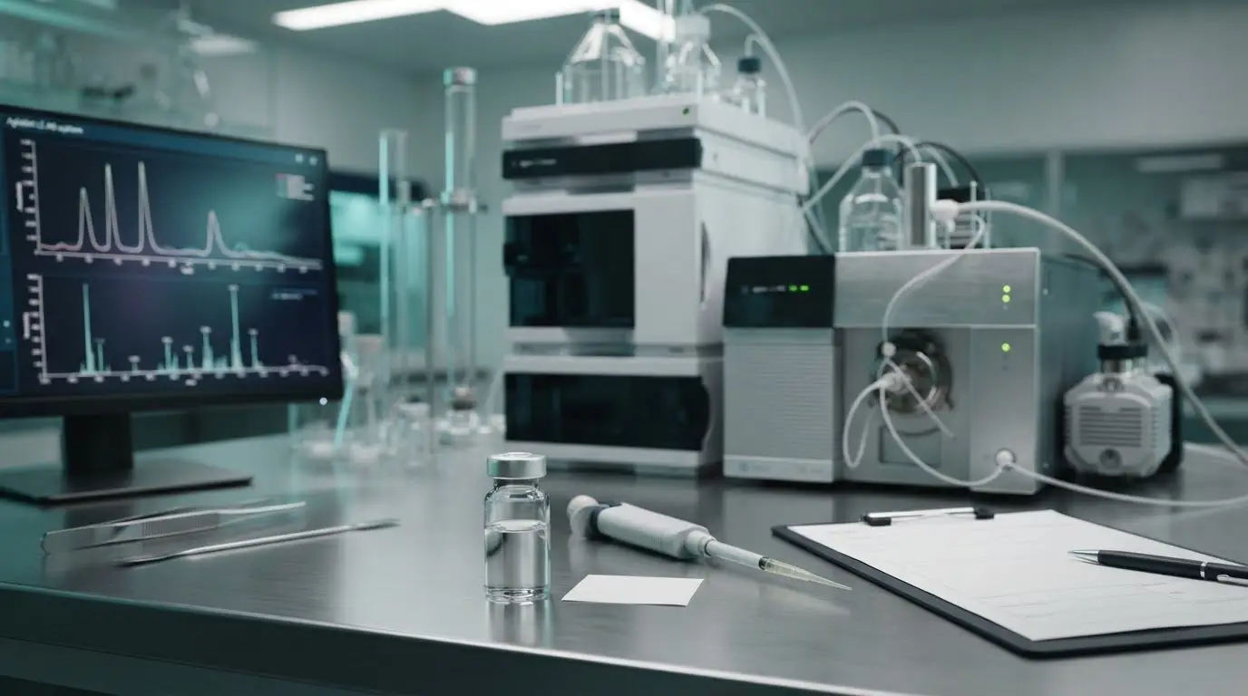
Analytical verification begins with reverse-phase HPLC chromatograms. Analysts calculate percentage purity via peak area normalization, measuring the primary complex peak area against the total integrated signal. A quality profile shows a sharp, symmetrical elution peak. Early-eluting minor peaks indicate truncated peptide fragments or free copper salt, while late-eluting spikes reveal hydrophobically shifted synthesis artifacts. LC-MS spectral data confirms molecular mass, displaying the characteristic ionized m/z peaks for the intact complex around 467.02 Da without uncomplexed peptide signals at 403.93 Da.
Solara Compounds Quality Standards for Laboratory Buyers
Solara Compounds establishes an uncompromising standard for transparent analytical verification in research peptide procurement. Every batch of Solara Compounds GHK-Cu (supplied in lyophilized 50mg and 100mg formats) is manufactured via solid-phase peptide synthesis in ISO 7 cleanroom environments. To remove bias and ensure batch-to-batch reproducibility, every production lot undergoes independent third-party analytical testing by Kovera Labs, confirming HPLC purity exceeding 98% alongside exact mass confirmation.
Transparency remains central to reliable science. Researchers can examine batch documentation directly through the public Solara Compounds COA database before placing an order. All items supplied through solaracompounds.com are strictly for laboratory research only, never for human or animal consumption. Lab buyers managing high-throughput cell trials also benefit from fast domestic fulfillment across the United States, including free shipping on qualifying orders over $200.
Advancing Precision in Dermal In Vitro Modeling
Translating the biological signaling of copper tripeptides into reliable assay data requires separating commercial marketing from empirical cell science. Success in GHK-Cu skin biology research depends on three fundamentals: establishing physiological concentrations in validated cell models, safeguarding aqueous stability against pH drift and chelator dissociation, and verifying analytical purity before running assays.
Experimental consistency starts with verified chemistry. Solara Compounds supports reproducible testing by subjecting every lot to independent third-party batch testing by Kovera Labs. This process guarantees strict >98% HPLC purity and confirms intact copper chelation via mass spectrometry, supported by publicly accessible batch-specific COAs. All compounds are provided strictly for laboratory research only, never for human or animal use.
Equip your benchtop with reliable reagents that keep your cellular assays consistent. To review analytical documentation and select verified materials for your laboratory, view the current research catalog at solaracompounds.com.
Frequently Asked Questions
What is the molar binding ratio of copper to the GHK tripeptide in laboratory compounds?
The stoichiometric molar binding ratio of copper to the GHK tripeptide is exactly 1:1. Each molecule of glycyl-L-histidyl-L-lysine coordinates with a single divalent copper ion [Cu(II)] in a square-planar geometry. Research preparations must maintain this equimolar ratio. Excess unchelated copper ions cause non-specific cytotoxicity in cell models, while an excess of uncomplexed peptide alters baseline receptor binding dynamics in GHK-Cu skin biology research.
How does GHK-Cu differ from unchelated GHK peptide in cell culture assays?
Unchelated GHK lacks the coordinated Cu(II) ion, which significantly alters its biological activity in vitro. While plain GHK exhibits weak carrier properties, GHK-Cu actively serves as a copper chaperone, donating copper to copper-dependent enzymes like lysyl oxidase. In comparative fibroblast culture models, the chelated complex demonstrates substantially higher gene-modulating activity and extracellular matrix signaling than the free peptide base alone.
Can GHK-Cu be reconstituted in standard phosphate-buffered saline (PBS)?
Yes, GHK-Cu dissolves readily in sterile, isotonic phosphate-buffered saline at physiological pH (7.2 to 7.4). However, PBS solutions lack antimicrobial preservatives and antioxidant stabilizers. Researchers conducting cellular assays should prepare PBS working solutions immediately before application and use them within 24 hours. Prolonged storage in unpreserved saline risks ambient copper oxidation, peptide cleavage, and microbial growth that can invalidate sensitive cell culture trials.
What analytical methods are required to verify research-grade GHK-Cu purity?
Verifying research-grade GHK-Cu requires high-performance liquid chromatography (HPLC) combined with mass spectrometry (LC-MS or MALDI-TOF). Reverse-phase HPLC determines chemical purity, requiring a minimum benchmark of >98% through peak area integration. Mass spectrometry confirms the exact molecular mass (467.02 Da for the chelated complex) and verifies stoichiometric copper binding. Facilities in research hubs like Boston, San Diego, and Raleigh rely on third-party testing from labs like Kovera Labs to validate batch metrics.
What happens if GHK-Cu solutions are exposed to chelating agents like EDTA?
Strong synthetic chelators like EDTA possess a higher binding affinity for divalent copper than the GHK peptide. When exposed to EDTA, the copper ion dissociates from the tripeptide core almost immediately. This leaves the naked peptide backbone and generates free copper-EDTA complexes. In GHK-Cu skin biology research, this chemical stripping invalidates matrix remodeling assays and alters cellular uptake, making extensive cell washes essential after enzymatic harvesting.
How should lyophilized GHK-Cu be stored to ensure long-term stability?
Lyophilized GHK-Cu vials should remain desiccated and frozen at -20°C for routine storage, or at -80°C for multi-year preservation. Vials must stay sealed to protect the freeze-dried cake from ambient moisture, which induces slow hydrolytic degradation. Before reconstitution, laboratory buyers should allow vials to equilibrate to room temperature for 20 minutes to prevent condensation inside the container during handling.
Is GHK-Cu approved for human cosmetic application or clinical treatment?
No, GHK-Cu supplied by Solara Compounds is classified strictly as a Research Use Only (RUO) chemical complex. It doesn’t carry approval from the FDA, EMA, or other regulatory bodies for clinical therapy, medical treatment, veterinary use, or direct human application. Investigators across scientific centers from San Francisco to New York procure this material exclusively for in vitro modeling, biochemical evaluation, and controlled laboratory experiments.